 aide
aide
Recherche - Valorisation
Photochemical analysis of structural transitions in DNA liquid crystals reveals differences in spatial structure of DNA molecules organized in liquid crystalline form
Brach, K., Hatakeyama, A., Nogues, C., Olesiak-Banska, J., Buckle, M., & Matczyszyn, K. (2018). Scientific reports, 8(1), 4528

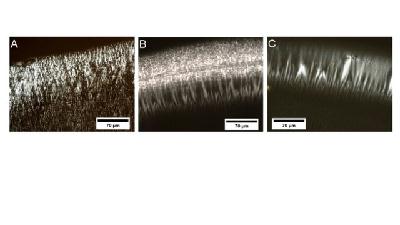
Polarized light microscopy observation of Liquid Crystal DNA. DNA LC texture (A) at the perimeter of a dried drop for DNA in water; (B) at the perimeter of a dried drop for DNA in 10 mM Tris at pH 7.2 and (C) at the perimeter of a dried drop for DNA in 10
The anisotropic shape of DNA molecules allows them to form lyotropic liquid crystals (LCs) at high concentrations. This liquid crystalline arrangement is also found in vivo (e.g., in bacteriophage capsids, bacteria or human sperm nuclei). However, the role of DNA liquid crystalline organization in living organisms still remains an open question. Here we show that in vitro, the DNA spatial structure is significantly changed in mesophases compared to non-organized DNA molecules. DNA LCs were prepared from pBluescript SK (pBSK) plasmid DNA and investigated by photochemical analysis of structural transitions (PhAST). We reveal significant differences in the probability of UV-induced pyrimidine dimer photoproduct formation at multiple loci on the DNA indicative of changes in major groove architecture.
 aide
aide
